Биоструктурные исследования, в частности — анализ белков, требуют точного понимания их размера, формы, агрегации и стабильности для обеспечения надежных результатов и оптимизации экспериментальных процессов. Из статьи наших партнеров XENOCS вы узнаете, как лабораторный метод SEC-SAXS позволяет проводить разделение и структурную характеризацию белков в режиме реального времени, предоставляя детальную информацию о мономерах, димерах и агрегатах. Такой подход оптимизирует ваши исследования и предоставляет важные данные для разработки биопрепаратов. Статья на английском языке
Laboratory-based size-exclusion chromatography SAXS used for advanced characterization of protein solutions.
The structural and conformational stability of biological samples presents significant challenges due to their complex structures and sensitivity to environmental conditions such as pH, temperature, high concentration, and the presence of excipients [1]. These challenges include maintaining structural integrity, preventing nonspecific aggregation, minimizing oligomerization and degradation, and ensuring chemical and physical stability.
Small Angle X-ray Scattering (SAXS) is a well-established technique that provides detailed information on the size, shape, structure, stability, flexibility, oligomeric state, folded/unfolded states, and interparticle interactions of samples in a near-native environment, regardless of size [1,2]. SAXS intensity profiles are influenced by the contributions of each particle in the sample, weighted by their X-ray contrast and volume fraction. Consequently, sample heterogeneity, such as the presence of oligomers and aggregates, can add complexity to SAXS analyses, potentially influencing data quality and the accuracy of structural interpretations. Overcoming these challenges requires cutting-edge analytical techniques, and the integration of size-exclusion chromatography (SEC) with SAXS has proven to be an effective solution [3]. SEC-SAXS combines the separation capabilities of SEC with the detailed structural analysis of SAXS, delivering real-time, high-resolution insights into macromolecular behavior.
To illustrate the capabilities of laboratory-based SEC-SAXS, we present a detailed analysis of bovine serum albumin (BSA). In a typical SEC-SAXS measurement, a BSA sample is first passed through the SEC column, which separates monomeric BSA from aggregates, oligomers and degradation products [2-4]. The fractions eluted from the column are directly passed through the X-ray beam, and the scattering intensity is measured in timed frames.
Measurements
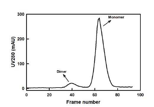
The BSA sample, dissolved in HEPES buffer, was injected into a sizeexclusion chromatography column using an AKTA Go system. The SEC column was connected in-line with a Xeuss laboratory SAXS instrument. Approximately 300 μl of 10 mg/ml BSA sample was loaded on the column. The flow rate was set to 0.5 ml/min and decreased to 0.1 ml/min when the protein eluted from the column, in order to ensure long enough exposure times. SAXS data were collected at 25 °C with 30-second exposure times and a sample–detector distance of 600 mm. The sample concentration was monitored directly in the SAXS exposed volume using UV absorbance at 280 nm during the measurements (as presented in Figure 1). The Xenocs XSACT Pro software was used for data reduction, analysis and visualization.
Figure 1. SEC profile of BSA using a Superdex 200 increase
10/300 GL column with a bed volume of 24 ml. The UV
absorbance was measured directly by the Xeuss laboratory
SAXS instrument.
Results
The UV profile showed two well resolved peaks labelled as the dimer and the monomer peaks in Figure 1, indicating that BSA predominantly consists of monomers with a small proportion of dimers. The individual buffer-subtracted frames corresponding to the monomer and dimer peaks were analyzed for radius of gyration (Rg) and maximum dimension (Dmax) (Fig. 2). The analysis revealed an Rg of 2.77 Ѓ} 0.04 nm and a Dmax of 8.5 nm for the monomer, and an Rg of 3.83 Ѓ} 0.8 nm and a Dmax of 13 nm for the dimer, both in good agreement with previously reported data [3-5].
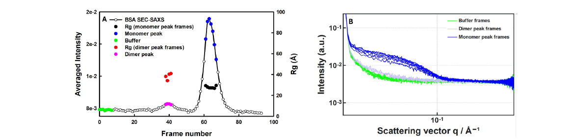
Figure 2. Selection of sample and buffer frames for analyzing SEC-SAXS data from BSA samples. (A) A
chromatogram (produced by CHROMIXS [6]) showing the averaged SAXS intensity and Rg values of monomer
and dimer peaks as a function of frame index. (B) Individual unsubtracted 30 s frames for buffer, monomer and
dimer fractions.
SAXS curve fitting analysis of the BSA crystal structure and dimer model yielded an excellent fit to the observed SAXS data (Fig.3). The monomer model gave an Rg value of 2.78 nm, Dmax of 8.5 nm and a χ2 of 0.8, while the dimer model exhibited an Rg value of 3.9 nm, Dmax of 14 nm and a χ2 of 0.7, validating the observed SAXS data.
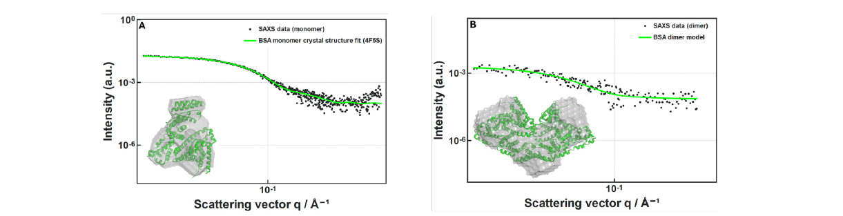
Figure 3. (A) Fit of the BSA monomer crystal structure (green line) to the SAXS data averaged from 7 frames (black
dots), (B) Fit of the BSA dimer crystal structure (green line) to the SAXS data averaged from 4 frames (black dots).
The insets show ab initio models generated by DAMMIF [7] based on the SAXS data, overlayed with the respective
crystal structures. The crystal structures were taken from PDB ID: 4F5S.
Conclusion
By enabling SEC-SAXS data collection with an in-house SAXS instrument, researchers can more efficiently analyze transient, polydisperse and unstable biomacromolecules in solution. This method eliminates the need for separate chromatography and purification steps, streamlining the research processes. Using BSA as an example, this study highlights the practical advantages of SEC-SAXS in laboratory settings, emphasizing its valuable role in advancing biologics research and development.
Author: Sanaullah Khan
Measurements and Data Analysis: Sanaullah Khan
Scientific reviewers: Soren Skou, Al Kikhney, Ana-Elena Tutueanu.
References
[1] A. Y. Xu, M. A. Blanco, M. M. Castellanos, C. W. Meuse, K. Mattison, et al., Role of Domain–Domain Interactions on the Self-Association and Physical Stability of
Monoclonal Antibodies: Effect of pH and Salt, J. Phys. Chem. B 127, 8344 (2023). DOI: 10.1021/acs.jpcb.3c03928.
[2] S. Da Vela and D. I. Svergun, Methods, development and applications of small-angle X-ray scattering to characterize biological macromolecules in solution,
Current Research in Structural Biology 2, 164 (2020). DOI: 10.1016/j.crstbi.2020.08.004.
[3] M. A. Graewert, S. Da Vela, T. W. Grawert, D. S. Molodenskiy, C. E. Blanchet, et al., Adding Size Exclusion Chromatography (SEC) and Light Scattering (LS)
Devices to Obtain High-Quality Small Angle X-Ray Scattering (SAXS) Data, Crystals 10, 11 (2020). DOI: 10.3390/cryst10110975.
[4] S. Bucciarelli, S. R. Midtgaard, M. Nors Pedersen, S. Skou, L. Arleth, and B. Vestergaard, Size-exclusion chromatography small-angle X-ray scattering of water
soluble proteins on a laboratory instrument, J Appl Cryst 51, 1623 (2018). DOI: 10.1107/S1600576718014462.
[5] C. M. Jeffries, M. A. Graewert, C. E. Blanchet, D. B. Langley, A. E. Whitten, and D. I. Svergun, Preparing monodisperse macromolecular samples for successful
biological small-angle X-ray and neutron-scattering experiments, Nat Protoc 11, 2122 (2016). DOI: 10.1038/nprot.2016.113.
[6] A. Panjkovich and D. I. Svergun, CHROMIXS: automatic and interactive analysis of chromatography-coupled small-angle X-ray scattering data, Bioinformatics
34, 1944 (2018). DOI: 10.1093/bioinformatics/btx846.
[7] D. Franke and D. I. Svergun, DAMMIF, a program for rapid ab-initio shape determination in small-angle scattering, J Appl Cryst 42, 342 (2009). DOI: 10.1107/